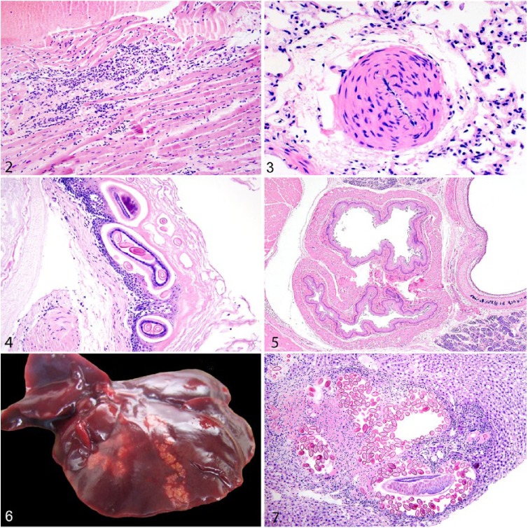
Figures 2–7.
Microscopic lesions in urban Norway rats. Figure 2. Lymphocytic myocarditis, myocyte necrosis, and fibrosis indicative of cardiomyopathy. H&E. Figure 3. Medial hypertrophy of pulmonary blood vessels featuring thickening of the tunica media by disorganized smooth muscle cells. H&E. Figure 4. Eucoleus sp. adults and eggs in the non-glandular gastric mucosa in association with hyperkeratosis and mucosal hyperplasia. H&E. Figure 5. Two distinct esophageal lumens (double esophagus) are adjacent to the trachea. H&E. Figure 6. Multiple tortuous tan parasite tracts caused by Capillaria hepatica on the liver surface. Figure 7. An adult C. hepatica in oblique section and eggs efface hepatocytes and are surrounded by granulomatous inflammation and fibrosis in the liver parenchyma. H&E.

