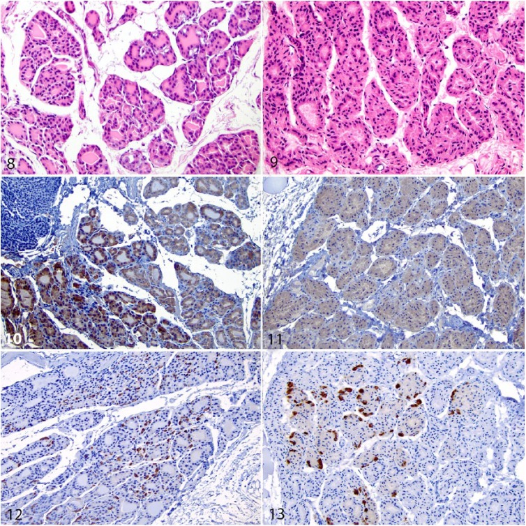
Figures 8–13.
Normal and diffuse thyroid follicular hyperplasia in the thyroid gland of urban Norway rats. Figure 8. Normal thyroid follicular cells are low cuboidal and surround follicles that contain abundant, homogeneous, eosinophilic colloid. H&E. Figure 9. Affected thyroid follicular cells are cuboidal to low columnar and surround follicles that lack colloid. H&E. Figure 10. Normal immunostaining that is intense and intracytoplasmic within follicular cells. Immunohistochemistry (IHC) for thyroglobulin. Figure 11. Within affected hyperplastic cells, immunostaining is weak, diffuse, and intracytoplasmic, consistent with follicular cells. IHC for thyroglobulin. Figures 12, 13. Scattered normal interstitial C cells have intense cytoplasmic immunostaining, whereas hyperplastic follicular cells are negative. IHC for calcitonin.

