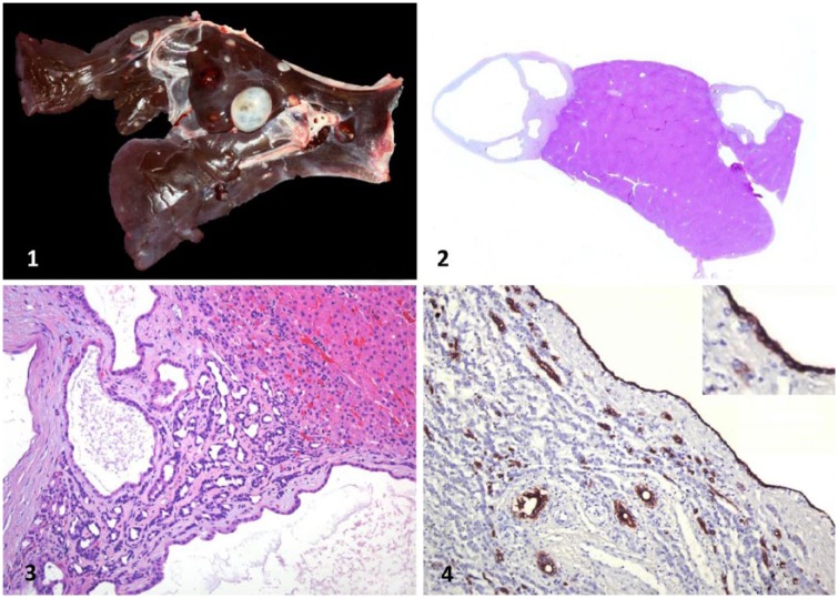
Figures 1–4.
Polycystic liver in an adult llama (Lama glama). Figure 1. Liver with multiple round-to-oval biliary cysts of various sizes. The cysts are surrounded by a capsule of fibrous collagenous tissue. Figure 2. Sub-gross photograph of liver with multiple cysts. H&E. Figure 3. Hepatic cysts composed of a single cuboidal-to-columnar epithelium surrounded by a thin fibrous capsule. H&E. Figure 4. Intense intracytoplasmic immunohistochemical reactivity of cystic and bile duct epithelium for cytokeratin 19. Inset: higher magnification of cyst epithelium.

