Figures 1–4.
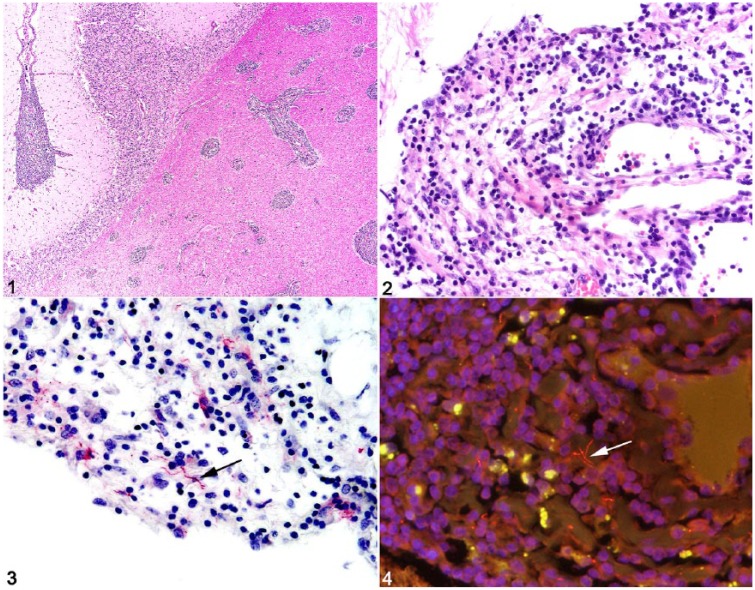
Neuroborreliosis in a horse with common variable immunodeficiency. Figure 1. Cerebral Virchow–Robin spaces are expanded by large numbers of lymphocytes and histiocytes. Figure 2. Lymphohistiocytic and neutrophilic cerebral meningitis. Figure 3. Rare immunoreactive Borrelia burgdorferi spirochetes within areas of meningitis (arrow). Figure 4. Probe-positive spirochetes within the cerebrum (arrow).

