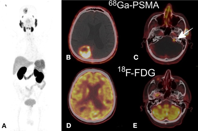
Figure 4.

(A) Maximum intensity projection 68Ga-PSMA (MIP), (B,C) 68Ga-PSMA axial PET/CT fusion demonstrating a non-homogeneous uptake in the right parietal mass (B) and a lower uptake in the left auditory neuroma (C), (D,E) 18F-FDG axial PET/CT fusion showing an increased uptake comparable to that in the gray matter in the parietal tumor (D) and no uptake in the neuroma (E). This research was originally published in Kunikowska et al. (186).
