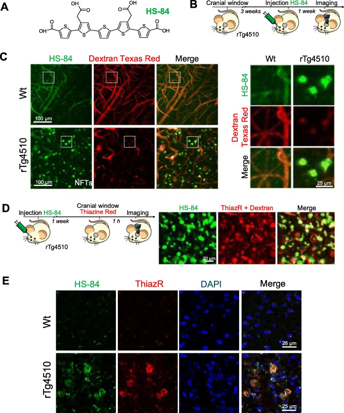
Fig. 1.
HS-84 binds to NFTs in the rTg4510 Tg mouse and can be detected with multiphoton microscopy. a. HS-84 molecule. b. Experimental procedure to characterize HS-84 in the mouse brain in vivo. rTg4510 Tg mice and Wt littermates carrying a cranial window were injected with HS-84 via retro-orbital and subjected to intravital multiphoton microscopy. c. In vivo multiphoton microscopy images of HS-84 in Wt (top) and rTg4510 Tg mouse (bottom). Pictures show HS-84 (green), Dextran Texas Red (70,000 Da MW, red), and merge of both channels. Insets are shown on the right. Representative from n = 3 Wt and n = 6 rTg4510 Tg mice. d. In vivo validation of HS-84 labelling NFT. Left, experimental procedure to validate HS-84 in the mouse brain in vivo. rTg4510 Tg mice were injected with HS-84. 1 week later, Thiazine Red was directly applied for 1 h onto the brain after skull and dura removal. A 8 mm cranial window was implanted and the mouse was subjected to multiphoton microscopy. Right, HS-84 colocalizes with Thiazine Red. Representative of n = 2 rTg4510 Tg mice. Scale bar represents 20 μm. e. Post-mortem validation of NFTs labelled with HS-84 in the mouse brain. HS-84 was injected via retro-orbital and the mice were euthanized 1 week later. Brains were sliced in a cryostat and incubated with 0.005% Thiazine Red in EtOH. Colocalization of both dyes can be appreciated. Scale bar represents 25 μm and applies to all pictures

