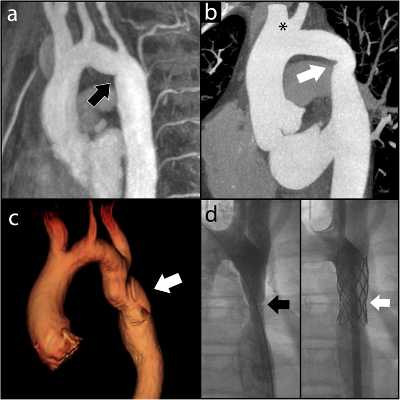
Fig. 4.

Examples of CoA repair. a Oblique sagittal maximum intensity projection reconstruction of MRA performed for a patient who underwent end-to-end anastomotic CoA repair. A mild fold is demonstrated at the CoA repair site at the aortic isthmus (arrow). b Oblique sagittal maximum intensity projection reconstruction CT angiogram for patient with subclavian flap CoA repair. There is absence of the proximal left subclavian artery with mild narrowing at the site of coarctation repair in the distal arch (arrow). Note normal variant conjoint origin of the right brachiocephalic and left common carotid artery (asterisk). c 3D reconstruction MRA in a patient who underwent patch repair of significant CoA. The white arrow indicates a pseudoaneurysm in the proximal descending aorta, which developed at the site of repair. d Fluoroscopic images of CoA stent procedure. Left panel shows CoA in the proximal descending aorta (black arrow). Right panel shows successful stent implantation with improved patency of the proximal descending aorta (white arrow)
