
Figure 3.
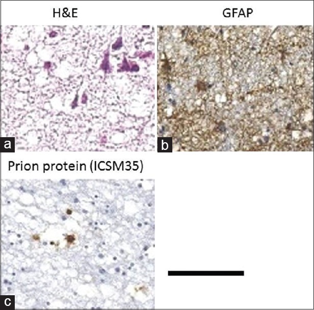
Neuropathology: (a) Grey matter (H and E): Severe spongiform changes and neuronal loss; (b) Grey matter: (GFAP stain) gliosis along the cortex; (c) Caudate nucleus: Immunostaining showing the position of prion protein. Scale bar: 50 μm – A, 200 μm – B and C