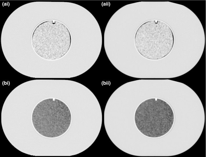
Figure 2.

CT scans of the radiomics phantom used in this study. To the left (i‐a and i‐b) are the scans referred to first time point and to the right (ii‐a and ii‐b) is the second time point in this study. Top two scans (i‐a and ii‐a) are the cartridges containing rubber and the two bottom ones (i‐b and ii‐b) have cartridges with the material cork.
