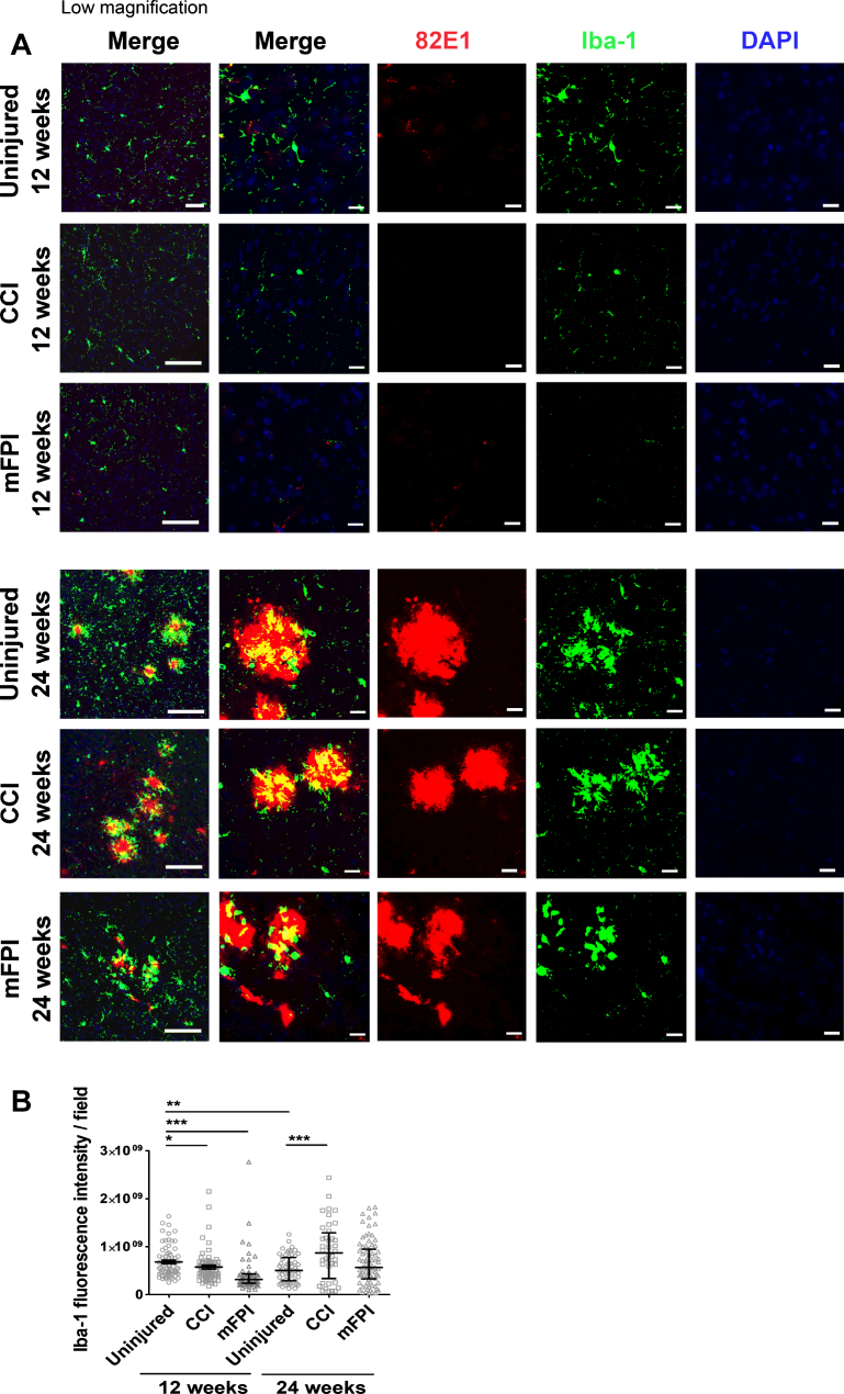
Fig. 5.
Induced number of activated microglia 24 weeks after CCI. Immunostainings with antibodies specific for Iba-1 (green) and the Aβ antibody 82E1 (red) demonstrated that microglia were found in close proximity to the plaques (A). At 12-weeks post-TBI there was a sparse expression of Iba-1 in both uninjured and injured mice. There was significantly less Iba-1 expression in mice that received CCI or mFPI at this time point, compared to uninjured controls. 24 weeks after TBI there was a two-fold increase of Iba-1 reactivity in mice that received CCI, compared to uninjured tg-ArcSwe mice (B). The data was analyzed by nonparametric Mann Whitney test and the graph displays mean±SEM. Scale bars: Low magnification = 20μm, high magnification = 50μm. *p < 0.05, **p < 0.01, ***p < 0.001.

