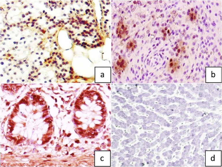
Figure 4.
Parafibromin immunohistochemistry (magnification ×400) showing (a) diffuse positivity in normal parathyroid tissue as compared with (b) sparse nuclear positivity in index parathyroid carcinoma. Representative images of (c) positive control (colonic mucosa) and (d) negative control (hepatocytes).

