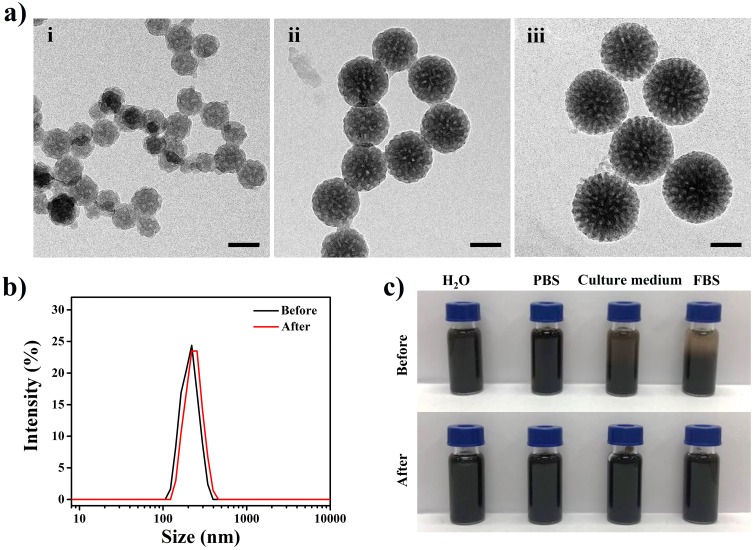
Figure 1.
(a) TEM images of MPDA nanoparticles: (i) ~80 nm, (ii) ~140 nm, and (iii) ~190 nm. The scale bars are 100 nm. (b) Hydrodynamic diameters of MPDA (~190 nm) before and after PEG modified. (c) Photographs of MPDA before and after PEG modified in different media (water, PBS, RPMI-1640 medium (containing 10% FBS), FBS) for 4 hrs.
Abbreviations: TEM, transmission electron microscope; MPDA, mesoporous polydopamine; PEG, polyethylene glycol; PBS, phosphate buffer saline; RPMI, Roswell Park Memorial Institute; FBS, fetal calf serum.

