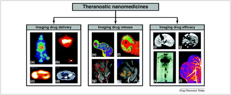
FIGURE 2.
Theranostic drug delivery system (DDS): is a new non-invasive imaging technique to check the ability of the pharmaceutical formulation to deliver the drug (a–d), to control drug release (e–h) and to determine the impact of this formulation on the drug efficacy (i–l).
(a) EPR induced accumulation of iodine −131 labeled HPMA copolymer in the Dunning AT1 animal model.
(b–d) Different imaging modalities such as gamma camera, SPECT, and CT are used to monitor the delivery of galactosamine targeted HPMA copolymer containing DOX and labeled with iodine −123 to liver cancer.
(e and f) MR was used as a tool to determine the percent of released DOX from the temperature-sensitive liposome (TSL) encapsulating both manganeses as MRI contrasting agent and DOX as a therapeutic agent.
(g and h) PLGA nanoparticle with Gd-DTPA, SPIO, and 5-FU core was used to create multiple contrasting signals in the tumor and give an idea about PLGA dissociation.
(i and j) Gd-labeled polypropylene diaminobutane dendrimers showed high affinity for liver cancer and metastatic lesions.
(k) Indium-111 labeled PEGylated liposomes demonstrated the ability of stealth NPs to bypass capturing by RES and accumulate in the tumor side in patients with Kaposi sarcoma.
(l) Gadolinium labeled HPMA copolymer showed a marked affinity and high anticancer potency in Dunning AT1 animal model. The images are adapted with permission from Ref. [35].

