
Figure 7.
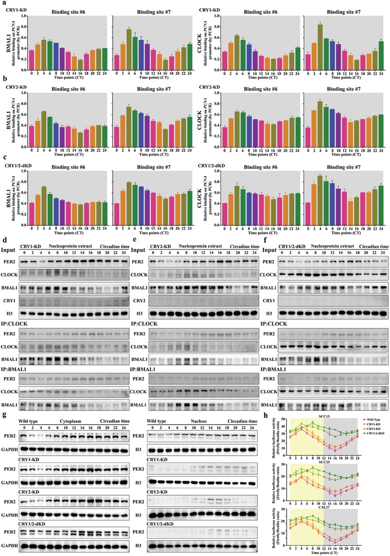
The functional process by which PER2 inhibits PCNA expression is dependent on CRY1/2. a–c) CLOCK and BMAL1 associate with PCNA promoter at predicted binding site #6 and #7 in CRY1 knockdown CAL27 cells, CRY2 knockdown CAL27 cells, and CRY1/2 double‐knockdown CAL27 cells at indicated time points. ChIP assay was performed using anti‐BMAL1 or anti‐CLOCK antibodies, with anti‐IgG antibody as a negative control. d–f) Coimmunoprecipitation (Co‐IP) assay was performed in nucleoprotein extracts obtained across a circadian cycle with anti‐CLOCK, anti‐BMAL1 antibody, or IgG (served as negative control) and detected by Western blot analysis with anti‐PER2, anti‐CLOCK, anti‐BMAL1, anti‐CRY1, or anti‐CRY2 antibodies in CRY1 knockdown CAL27 cells, CRY2 knockdown CAL27 cells, and CRY1/2 double‐knockdown CAL27 cells. g) Nucleocytoplasmic separation and Western blot analysis of cellular localization of PER2 in wild‐type, CRY1‐knockdown, CRY2‐knockdown, and CRY1/2 double‐knockdown CAL27 cells at indicated time points. h) A diurnal luciferase reporter assay was performed to measure the transcriptional activities of wild‐type PCNA promoter in human OSCC cells with CRY1‐knockdown, CRY2‐knockdown, and CRY1/2 double‐knockdown at indicated time points. Data represent the mean ± SD of three independent experiments.