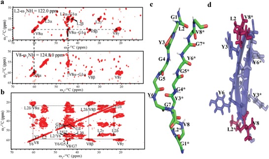
Figure 5.

Solid NMR characterization of 13C–15N labeled GV8 hydrogel. a) Strip plots of L2 and V8, 3D NCACX spectra of 13C–15N labeled GV8 peptide hydrogel displaying long‐range contact of residues. b) 2D 13C–13C DARR spectra with contact time of 50 ms showing long‐range dipolar contacts between L2 and V8 side chains (β, δ, γ). c) Representative structure of dimeric extended conformation of GV8 hydrogel. d) Side chain disposition representation of antiparallel β‐sheets of GV8 hydrogel displaying interchain connectivity between L2 and V8 residues (L2/V8* and L2*/V8).
