
Figure 4.
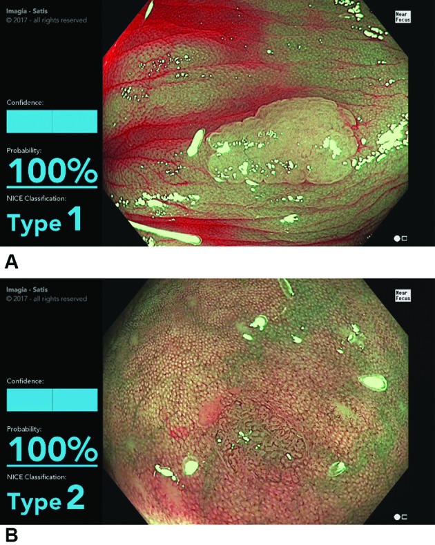
(A) Screen shot of the model during the evaluation of a NICE type 1 lesion (hyperplastic polyp). The display shows the type determined by the model (type 1) and the probability (100%). (B) Screen shot of the model in the evaluation of a NICE type 2 lesion (conventional adenoma). The display shows the type 2 determined by the model and the probability (100%) (see video). NICE, narrow band imaging International Colorectal Endoscopic.