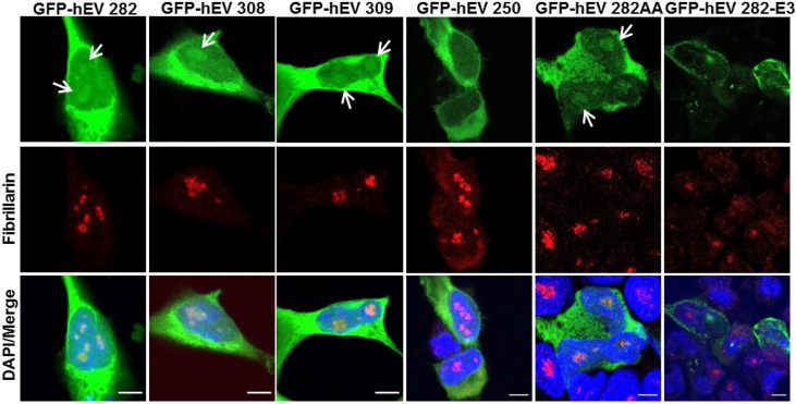
Fig 5. Subcellular distribution of ectopically expressed hENDOV proteins.
HEK 293T cells were transiently transfected for expression of GFP fused hENDOV isoforms (GFP-hEV 282, GFP-hEV 308, GFP-hEV 309, GFP-hEV 250, GFP-hEV 282AA and GFP-hEV 282-E3) (green). Cells were stained with a Fibrillarin antibody to visualize nucleoli (red). Nuclei are shown by DAPI staining (blue) and colocalization is shown in merged images. Arrows show dots representing nucleolar localization of GFP-hEV isoforms. Confocal images were acquired using an x40 oil objective, with 6–8x zoom (Leica SP8). Bar 5μm.

