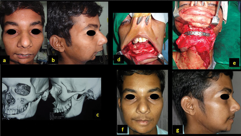
Fig. 2.

A 21-year-old man with previous reconstruction of symphysis with free fibula for benign lesion ( a , b ); CT scan showing the deformity with previous fibula ( c ). Reconstruction with osteocutaneous fibula ( d , e ). Postoperative outcome with dental rehabilitation ( f , g ).
