Figure 1.
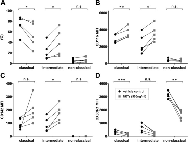
Stimulation of monocytes with NETs in vitro. (A) Monocyte subset shift as % of total monoctes, expression of (B) CD11b, (C) CD142 and (D) CX3CR1 of classical, intermediate and non-classical monocytes. Monocyte subsets in percent (%) or mean fluorescence intensity (MFI) levels are displayed. Monocytes were stimulated in whole blood from healthy donors (n = 5) with NETs derived from isolated neutrophils from a healthy donor (n = 1) or vehicle control for 60 minutes. *p < 0.05, **p < 0.01, ***p < 0.001.

