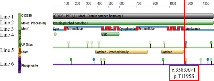
Figure 5.
The alignment of the PTCH1 amino acid sequence of reference and the biology effect of the mutated sites for posttranslational modification. The 1st line is the UniProt Knowledgebase (UniProtKB) sequence of human PTCH1 protein, entry number Q13635. The 2nd line is the full sequence of PTCH1, patched homolog 1. The structural motif of PTCH1 protein is drawn in line 3. The transmembrane helices are shown as red boxes, and the extracellular and intracellular domains are shown as blue lines. The 4th line is the sterol-sensing domain (SSD) for cholesterol signaling. The 5th line is the binding site for ligand proteins (patched and patched family proteins). The 6th line is the known sites of posttranslational modification. The green nodes are sites of phosphorylation, and the blue nodes are sites of methylation. c.3591C (phosphorylation of p.S1197 protein) is expressed as a green node. The mutation site c.3583A > T (p.T1195S in the protein) is marked with a red line and is located in the intracellular domain at the C-terminus of PTCH1 protein.

