Figure 1.
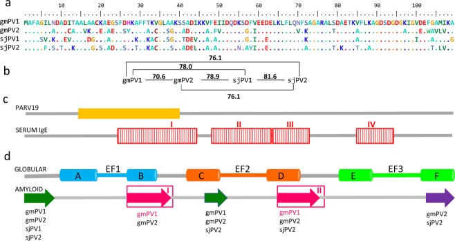
Sequences of the β-PV isoforms of Gadus morhua and Scomber japonicus. (a) Sequence alignment generated using BioEdit tools and their amino acid color table. (b) Pairwise identity calculated using BioEdit tools. (c) Relevant regions for PARV19 and serum IgE binding. The PARV19 epitope (orange rectangle) was predicted to include residues 13–3936. The displayed IgE binding epitopes (red rectangles) were determined using gmPV1-templated dodecapeptides25. (d) Segments involved in the globular and amyloid folds of β-PV. The globular fold contains three helix-loop-helix (AB, CD and EF) EF-hands, of which only CD and EF bind cations3,4,26,43. Regions assembling into amyloids were predicted by AmylPred2, and they are depicted by arrows. Regions known to form amyloids in gmPV1 are depicted with pink arrows25,30.

