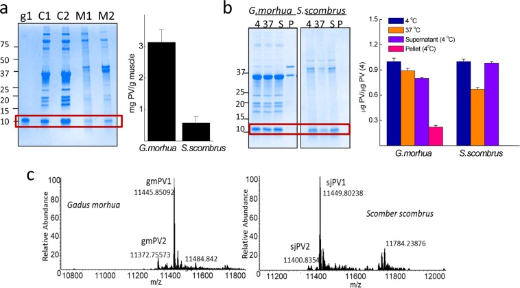
Figure 2.
Total PV content and relative abundance of β-PV isoforms in Gadus morhua and Scomber scombrus muscles. (a) Typical Coomasie Blue-stained SDS-PAGE gel of (C1, C2) Gadus morhua and (M1,M2) Scomber scombrus muscle extracts and the PV content estimated from monomer band quantification. The protein load per lane was 5 μg for the extracts and 0.5 μg for gmPV1, which was used as a control. Numbers on the right side indicate the molecular weights of markers in kDa. (b) SDS-PAGE analysis of the intrinsic proteolysis and solubility of PV in muscle extracts. Freshly prepared extracts were (4) stored at 4 °C, (37) heated for 15 min at 37 °C, cooled at 4 °C, and separated into soluble (S4) and insoluble (P4) fractions by ultracentrifugation. Numbers on the right side indicate the molecular weights of markers in kDa. (c) Mr determination for each of the different β-PV isoforms isolated from muscle extracts by FTICR-MS, considering the processing of M1 and the acetylation of A239,52. The original gels of panels a and b are displayed in supplementary Fig. S1.

