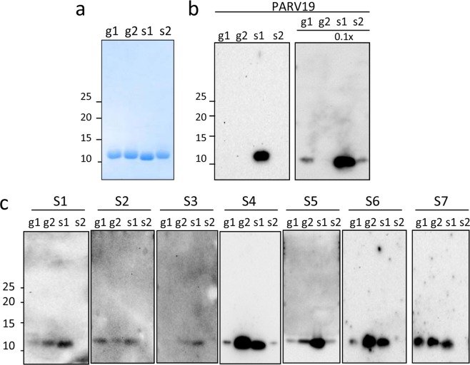
Figure 3.
Immunoreactivity of the denatured state of β-PV: linear IgE epitopes. (a) Coomassie blue-stained SDS-PAGE gel of β-PV isoforms. (b) Western blots of β-PV isoforms probed with PARV19. The relative loading amount is depicted at the top. (c) Western blots of β-PV isoforms probed with different fish-allergic patient sera for IgE binding. Displayed blots correspond to membranes developed independently. Numbers on the right side of gels and membranes in panels a and b correspond to the molecular weights of markers in kDa. Full-length gels and blots are displayed in supplementary Figs 2S and 3S.

