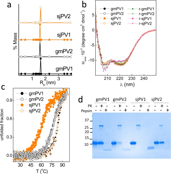
Figure 4.
Conformational properties of the Ca2+-bound globular folds of the distinct β-PV isoforms. (a) DLS analysis of the association state and hydrodynamic features. (b) Secondary structure probed by far-UV CD spectra. Spectra were recorded before (circles) and after heating (triangles) at 95 °C. (c) Thermal denaturation monitored changes in θ222 as a function of temperature. Symbol key: •, gmPV1; ◦, gmPV2;  , sjPV1;
  , sjPV2. (d) SDS-PAGE analysis of proteinase K and pepsin digestion products of β-PV isoforms. Digestions were performed using a 1/50 protease/β-PV ratio. The full-length gel is displayed in supplementary Fig. 4S.

