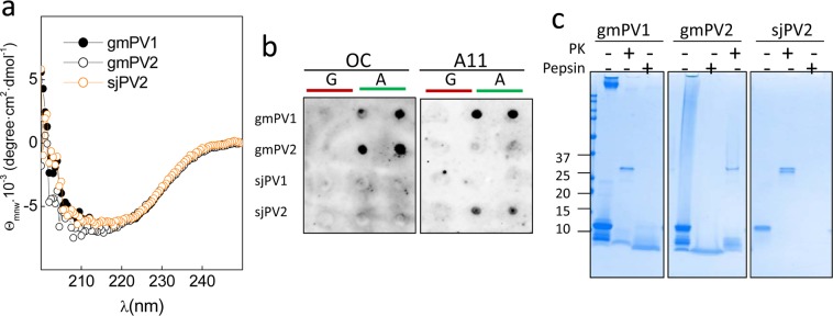
Figure 6.
Features of the amyloid aggregates formed by β-PV isoforms. (a) Far-UV CD spectra of the insoluble aggregates of gmPV1, gmPV2 and sjPV2. (b) Dot blot analysis of (A) β-PV aggregates using amyloid-specific anti-fibril (OC) and anti-oligomer (A11) antibodies. (G) Ca2+-bound chains were included as controls. (c) Coomassie blue-stained SDS-PAGE gel of the harsh proteinase K and pepsin digestions of gmPV1, gmPV2 and sjPV2 amyloid aggregates. The original membranes and gels of panels b and c are displayed in supplementary Fig. 6S.

