Fig. 2.
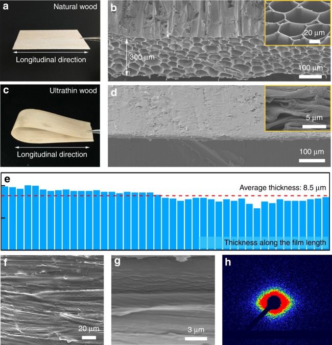
Morphological characterization of wood films. a Photograph of the rotary cut natural wood. b SEM image of the natural wood, with a thickness of 300 μm. Inset: top-view SEM image of the natural wood, showing its porous wood structure. c Photograph of the ultrathin wood. d SEM image of the ultrathin wood film, demonstrating its densified wood structure. Inset: Top-view SEM image of the ultrathin wood, revealing its collapsed wood cell walls. e The measured thickness of the ultrathin wood along its length at intervals of 5 μm, indicating uniform film thickness. f, g SEM images of the ultrathin wood, showing the aligned cellulose fibers. h Small-angle XRD pattern of the ultrathin wood, indicating the anisotropic alignment of the cellulose nanofibers

