Fig. 1.
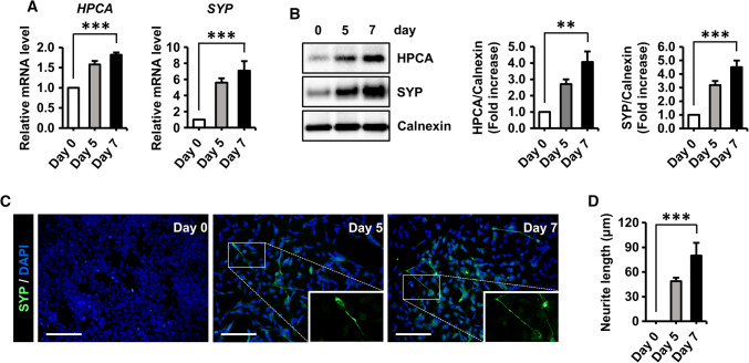
Differentiation induced expression of HPCA in SH-SY5Y cells. a, b SH-SY5Y cells were induced to differentiate by adding 50 μM all-trans retinoic acid (RA) for the indicated number of days. a The mRNA levels of the presynaptic marker, synaptophysin (SYP) and hippocalcin (HPCA), were determined by RT-qPCR. b Proteins were analyzed by western blotting with anti-HPCA, anti-SYP, and anti-calnexin antibodies. The graph shows mean densities as fold increases from five independent experiments (mean ± SEM). Band intensities were quantified using Quantity One® software. **P < 0.01, ***P < 0.001 compared with day 0 of differentiation. c Neuronal differentiation was induced by adding 50 μM RA for the indicated number of days. Cells were stained with DAPI (blue) and an anti-SYP antibody (green) to visualize neurite extensions. Scale bar, 100 μm. d Neurite lengths were measured in randomly selected areas in three independent experiments. ***P < 0.001 compared with day 0 of differentiation (mean ± SEM)

