Fig. 2.

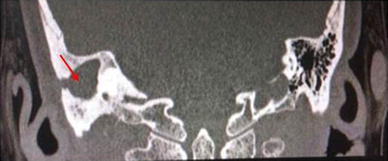
Coronal section of high resolution computed tomography (HRCT) scan of temporal bone, showing well pneumatized mastoid on left side and right mastoid showing secondary sclerosis with soft tissue density lesion (red arrow)

Coronal section of high resolution computed tomography (HRCT) scan of temporal bone, showing well pneumatized mastoid on left side and right mastoid showing secondary sclerosis with soft tissue density lesion (red arrow)