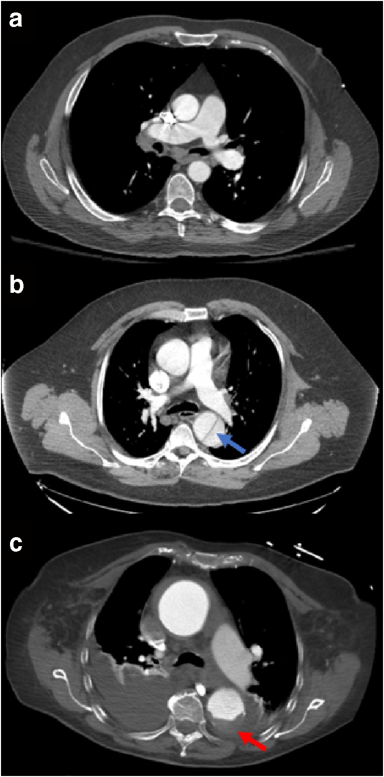
Fig. 1.

Axial post-contrast chest CT slices showing the aorta of 3 different patients. a A healthy aorta. b An aortic dissection (blue arrow). c A ruptured aorta (red arrow). Dark fluid is seen at the bottom of this image as blood from the rupture leaks into the surrounding tissues
