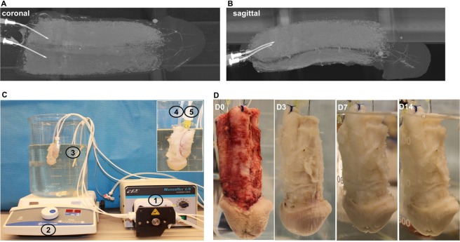
Figure 1.
Organ scaffold decellularization system. (A,B) Micro-CT radiographic images demonstrating the intricate vasculature via perfusion of contrast into the cavernosal arteries. (C) Perfusion system consisting of a (1) peristaltic pump, (2) magnetic stirrer plate, and a (3) 4 L glass container. The scaffold has been cannulated with a (4) Foley catheter in the urethra and (5) two angiocatheters in the cavernosal arteries. (D) Photographic representation of the decellularization process at 0, 3, 7, and 14 days, respectively.

