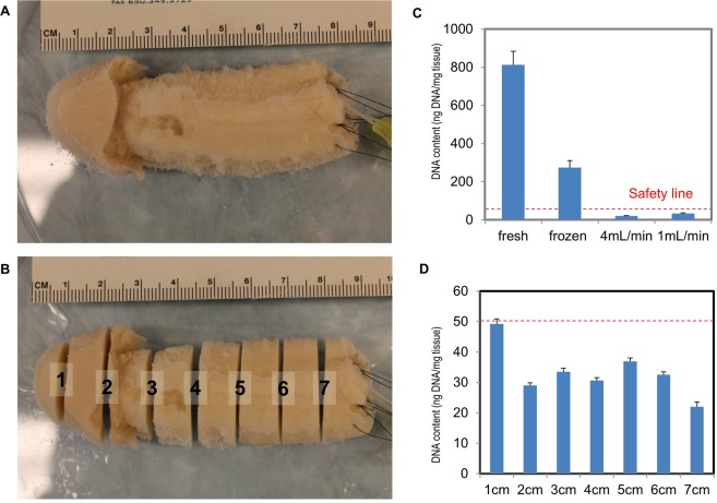
Figure 3.
DNA content quantification studies. A decellularized penile scaffold (A) before, and (B) after sectioning at 1 cm intervals. (C) Comparisons of fresh, frozen, and decellularized tissue at 4 mL/min and 1 mL/min perfusion rate for 2 weeks were quantified to evaluate DNA content. A red dotted-line indicates the threshold for an acceptable level of residual DNA which generates minimal to no post-implantation immunoresponse (50 ng DNA/mg tissue). (D) Sectioned slices along the organ scaffold are processed for DNA content quantification and to confirm a fully decellularized organ scaffold.

