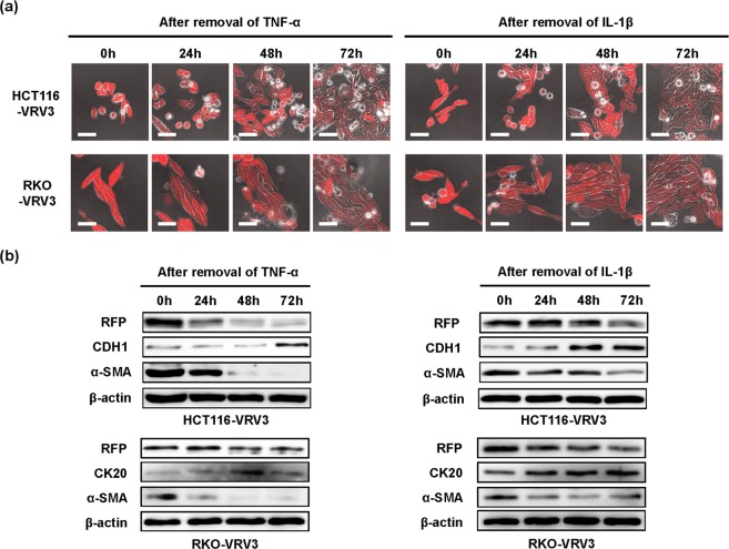
Figure 3.
Reversibility of TNF-α– and IL-1β–induced RFP expression and EMT phenotype. (a) Photographs of HCT116-VRV3 and RKO-VRV3 cells after removal of cytokines following treatment with TNF-α (20 ng/ml) or IL-1β (1 ng/ml) for 48 h. Scale bars: 50 μm. (b) Expression of RFP, epithelial markers (CDH1 and CK20), and a mesenchymal marker (α-SMA) in HCT116-VRV3 and RKO-VRV3 cells after removal of cytokines following treatment with TNF-α (20 ng/ml) or IL-1β (1 ng/ml) for 48 h. β-actin was used as a loading control.

