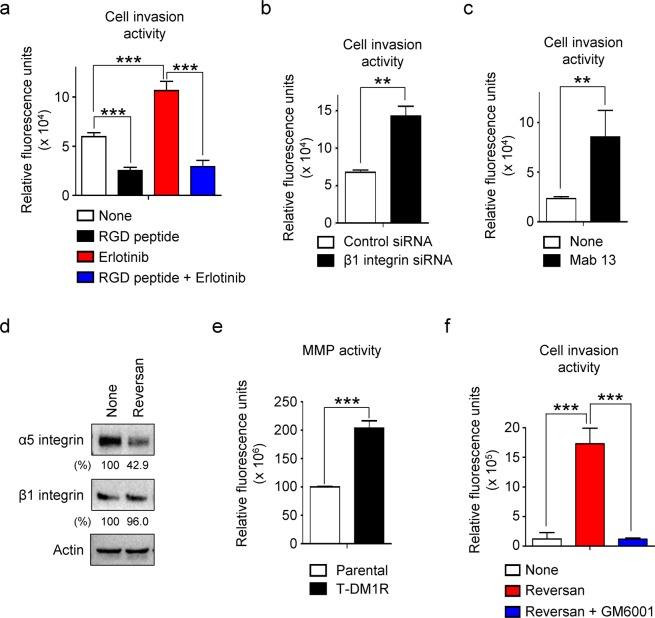
Figure 6.
Functional correlation between MRP1 and α5β1 integrin is conserved in breast cancer cells. (a) Cell invasion activity of MDA-MB-231 parental cells treated with either none or 5 µM erlotinib for 48 hrs in the media with or without RGD peptide. None vs. RGD peptide: p-value, 0.0001; none vs. erlotinib: p-value, 0.0007; erlotinib vs. RGD peptide + erlotinib: p-value, 0.0001. (b) Cell invasion activity of MDA-MB-231 parental cells treated with either control siRNA or β1 integrin siRNA for 48 hrs. Control siRNA vs. β1 integrin siRNA: p-value, 0.0003. (c) Cell invasion activity of MDA-MB-231 parental cells treated with an inhibitory antibody Mab 13 for 48 hrs. None vs. Mab 13: p-value, 0.0079. (d) The levels of α5 and β1 integrin expressions were evaluated by Western blot analysis in the WCL of T-DMR-MDA-MB-231 cells treated with either none or 2.5 µM Reversan for 48 hrs. (e) Total MMP activities were measured in the WCL of either MDA-MB-231 parental or T-DM1R cells. Parental vs. T-DM1R: p-value, <0.0001. (f) Cell invasion activity of MDA-MB-231-derived T-DM1R cells treated with either none or Reversan for 48 hrs with or without MMP inhibitor 50 nM GM6001. None vs. Reversan: p-value, 0.0003; Reversan vs. Reversan + GM6001: p-value, 0.0002.

