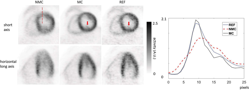
Figure 6:
NMC, MC and REF images for subject 1 (FDG) in short-axis and horizontal long-axis views. The REF images were obtained by reconstructing the data in the selected reference frame. Arrows indicate the papillary muscle, which is visible in both MC and REF, but not in NMC images. The profiles on the right were made along the dashed line shown on the short-axis image on the left.

