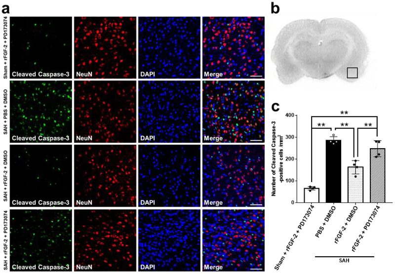
Fig. 9. Effects of pan-fibroblast growth factor receptor (FGFR) inhibitor on apoptotic marker after subarachnoid hemorrhage (SAH).
a Representative images demonstrating double immunofluorescent staining for cleaved caspase 3 and neuron-specific nuclear protein (NeuN) in the left temporal cerebral cortex at 24 h after modeling. Scale bar = 50 μm. b Representative image demonstrating the location of staining (small black box). c Quantitative analysis of number of cleaved caspase-3-positive cells per mm2 in the left cerebral cortex at 24 h after modeling. Data are expressed as mean ± standard deviation (n = 4 per group). *P < 0.05. DMSO, dimethyl sulfoxide; PBS, phosphate buffered saline; PD173074, pan-FGFR inhibitor; rFGF-2, recombinant fibroblast growth factor-2.

