
Figure 4. Untethered Heterochromatin in Lbr-Deficient Retinae.
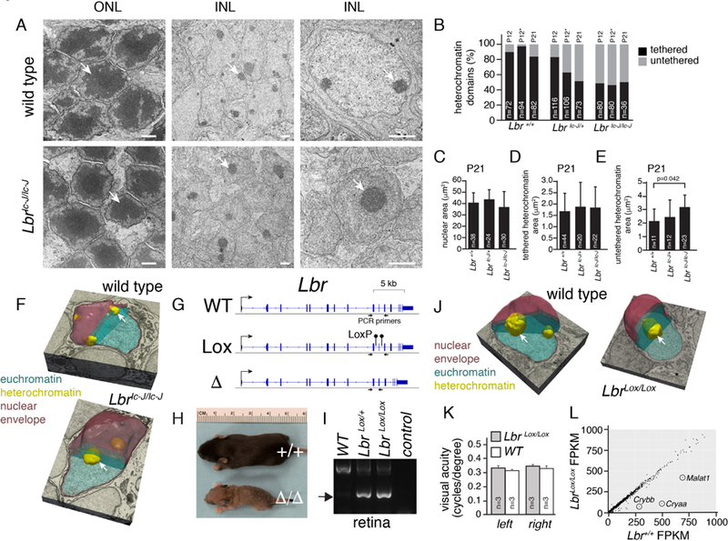
A) Electron micrographs of WT and Lbric-J/ic-J mouse retinae. Arrows indicate the heterochromatin domain in rods found in the outer nuclear layer (ONL) and neurons and glia found in the inner nuclear layer (INL). B) Stacked bar plot of the percentage of heterochromatin domains tethered or untethered to the nuclear lamina. The number of domains scored is indicated on each bar. The (*) indicates scoring on samples harvested at P12 and maintained in culture for 9 days. C-E) Bar plots of nuclear area (C), area of tethered heterochromatin domains (D), and untethered heterochromatin domains (E) from electron micrographs for Lbr+/+, Lbr+/ic-J, and Lbric-J/ic-J INL cells. F) 3D electron microscopy of bipolar nuclei in WT and Lbric-J/ic-J P21 retinae showing the difference in heterochromatin tethering (arrows). G) Genomic map of Lbr and location of PCR primers flanking the gRNA sequences used to produce LbrLox and Lbr∆ mouse strains with CRISPR-Cas9. H) Photo of P8 littermates with WT Lbr+/+ or deleted Lbr∆/∆ alleles. I) Photograph of an agarose gel with PCR products from retinal genomic DNA showing that the LbrLox allele can be recombined (arrow) in the Vsx2-Cre;LbrLox/+ or Vsx2-Cre;LbrLox/+ retina. J) 3D electron microscopy of bipolar nuclei in WT and LbrLox/Lox P21 retinae showing the difference in heterochromatin tethering (arrows). K) Barplot of visual acuity of WT and LbrLox/Lox adult mice measured on consequtive days over one week. Mean and standard deviation is plotted for 3 animals each. L) Scatterplot of gene expression for WT and LbrLox/Lox adult retina. Scale bars: 1 µm.