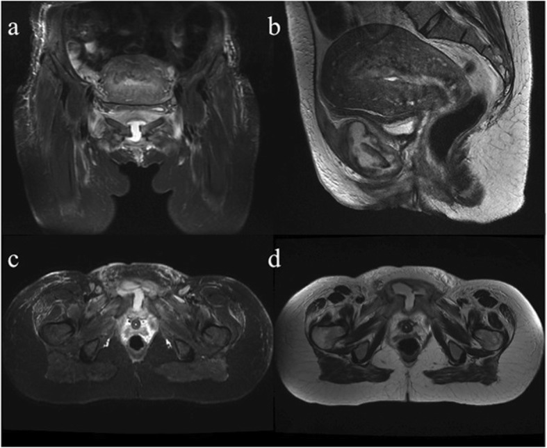
Fig. 2.

Pelvic magnetic resonance performed at day 8. Coronal short TI inversion recovery (STIR) (a), sagittal T2-weighted (b), axial STIR (c), and axial T2-weighted (d) images show pubic symphysis enlargement, abundant joint effusion with synovial thickening forming a pseudo-capsulated fluid collection within the symphysis, severe bone edema involving both pubic branches and edematous subcutaneous tissues
