Fig. 2.
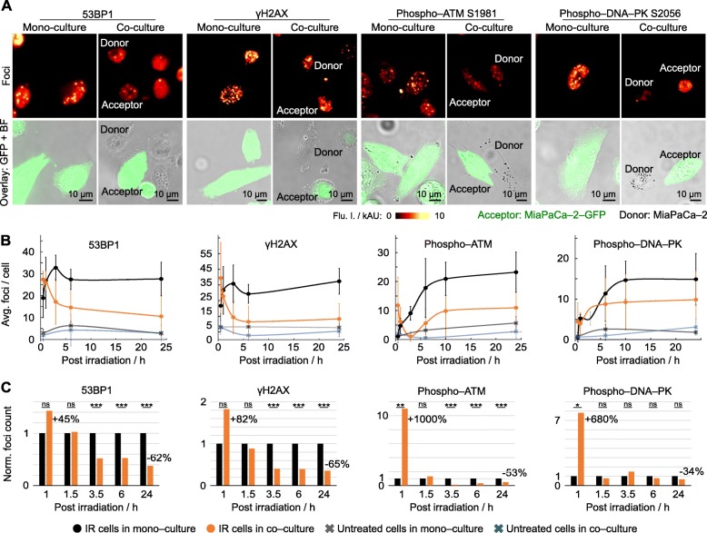
DNA repair is improved by presence of healthy cells. a Representative images of maximum intensity projections of indicated radiogenic foci in mono– and co–cultured MiaPaCa–2 cells at 24 h post irradiation (6 Gy x–rays). Foci visualized in acceptor (MiaPaCa–2–GFP; 6–Gy) and donor cells (unlabeled MiaPaCa–2; non–irradiated) by immunofluorescence staining. b and c Dynamic of radiogenic foci resolution over 24 h in absolute (b) and relative numbers (c) per irradiated cell (at least 50 cells analysed). Data represent mean ± SD (two–sided t–test; ns, not significant; *P < 0.05, **P < 0.01, ***P < 0.005)

