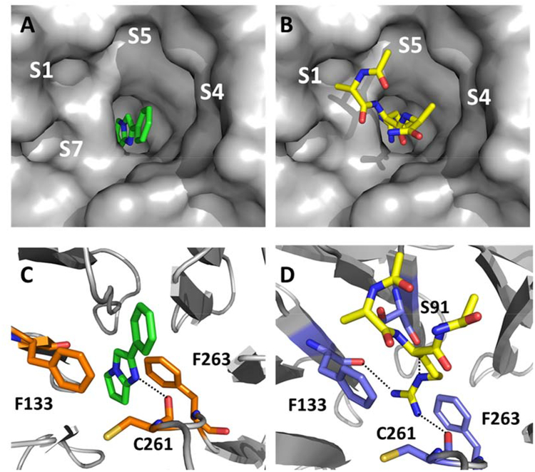
Figure 4.
X-ray co-crystal structure of fragment F-1 bound to WDR5 (PDB: 6D9X) and the reference MLL1 peptide bound to WDR5 (PDB: 3EG6) showing respective unoccupied and occupied pockets: A) F-1 left panel top view surface depiction; B) right panel top view MLL1 peptide surface depiction (only Ac-ARA-NH2 residues shown, yellow capped sticks); C) F-1 lower left panel, key residues and hydrogen bonding within S2 pocket (orange capped sticks); D) lower right panel MLL1 peptide (only Ac-ARA-NH2 shown) key residues and hydrogen bonding within S2 pocket (blue capped sticks).

