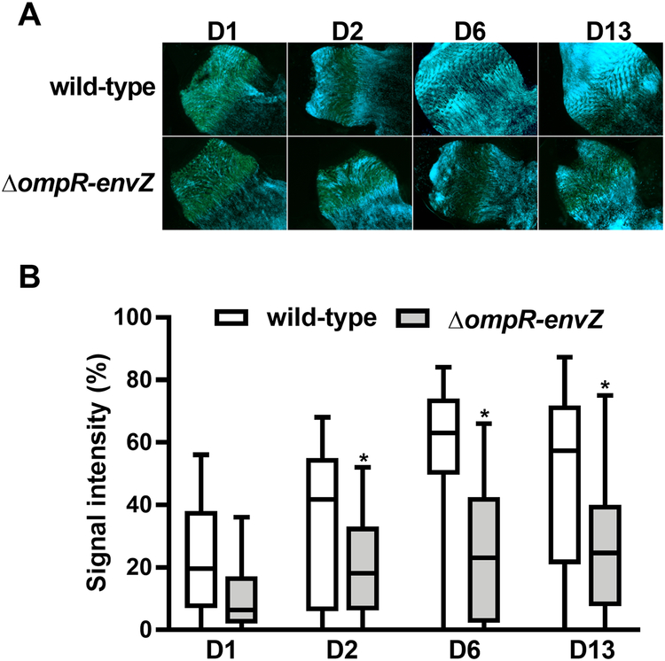
Figure 4. The Y. pestis OmpR-EnvZ system is required for massive colonization of the flea’s proventriculus.
(A) Representative fluorescence photos of the proventriculus colonized by the WT and the ΔompR-envZ mutant strain on different days (D) post-infection. The photos came from two independent experiments in which 17–20 proventriculi per strain were analyzed at each time point. (B) Box-and-whiskers (Tukey) represent the surface area occupied by bacteria in the proventriculus of 37 X. cheopis fleas that had fed on blood infected with the Y. pestis WT (white bars) and the ΔompR-envZ mutant strain (grey bars) and collected on different days (D) post-infection. The data represent the sum of 2 independent experiments in which 17–20 fleas were dissected (i.e. n= 37 per time point). The symbols indicate outliers. The signal intensities of the proventriculus infected with the WT vs. the ΔompR-envZ mutant strain differed significantly from the second day post-infection onwards (*, p<0.01 in a two-way analysis of variance with Sidak’s correction for multiple comparisons).

