
Figure 2:
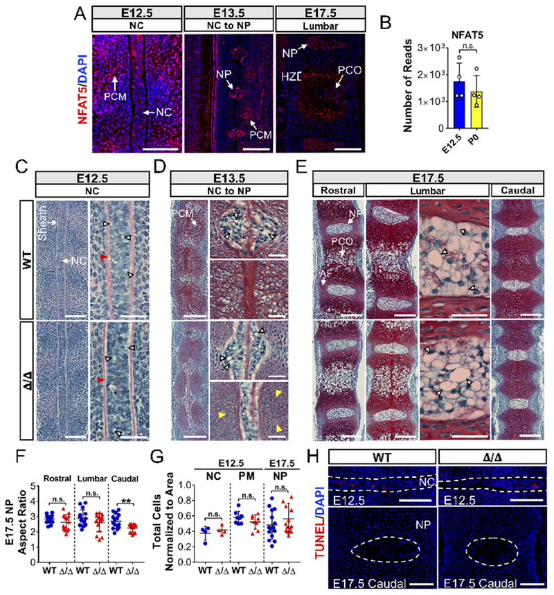
Loss of NFAT5 results in caudal discs with decreased aspect ratios. (A) Localization and expression of NFAT5 at E12.5, E13.5, and E17.5 by immunostaining. Scale bar = 100 μm. PCM: Perinotochordal condensing mesenchyme; NC: Notochord; NP: Nucleus pulposus; PCO: primary center of ossification; HZ: Hypertrophic zone. (B) NFAT5 mRNA expression by notochord cells at E12.5 and NP cells at P0 from deposited RNA-seq data (GSE100934). (C-E) Sagittal sections of E12.5, E13.5, and E17.5 embryos stained by Safranin-O/Fast Green/Hematoxylin showing notochord morphogenesis into NP at the thoracic, lumbar, and caudal levels. Scale bar = 50 μm. High magnification images showing intact perinotochordal sheath (red arrowheads), delayed enlargement of prevertebral cells at E13.5 (yellow arrowheads), and large intracellular vacuoles at all stages (white arrowheads) (Scale bar = 20 μm).(F) Aspect ratios of the caudal but not lumbar and rostral NP of NFAT5 null embryos were significantly smaller than WT embryos. (G, H) Changes in aspect ratio were not associated with decreased cell number per area or increased TUNEL positive cells. Scale bar = 100 μm. Quantitative measurements represent mean ± SD. n.s. = not significant; **, p ≤ 0.01.