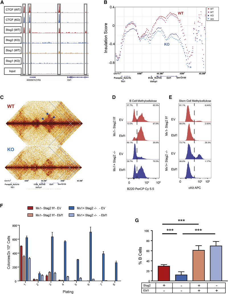
Fig 6. Induced Ebf1 expression rescues B cell development.
A) IGV track of the Ebf1 locus with Stag2 and Ctcf binding at 4 distinct sites lost in Stag2 KO and not bound by Stag1 either in WT or KO. B) ISC plotted across Ebf1 for Stag2 WT (n=2; shades of red) and KO (n=2; shades of blue) shows marked loss of insulation. C) Contact map of Ebf1 shows loss of cis-interaction at three loci (arrows). D-E) Lin− Stag2 WT and KO marrow were infected with retrovirus containing GFP-tagged empty vector or GFP-pCMV-Ebf1. GFP+ cells were plated in either B-cell colony methylcellulose or stem cell methylcellulose. Cells were harvested after 7 days and analyzed by flow cytometry for mature cell markers including (D) the B marker B220 or (E) stem cell markers including cKit. F) Cells plated in stem cell 10 methylcellulose were serially replated with exhaustion of Stag2 KO cell overexpressing Ebf1. G) GFP-tagged empty vector or GFP-pCMV-Ebf1 transduced Lin− cells were injected into lethally irradiated CD45.1 recipient mice. Leukocyte populations from the parent gate (GFP+, Cd45.2+, Cd45.1−) were analyzed by flow cytometry for percentage of mature B cells. Induced Ebf1 expression restored B cell population frequency from the Stag2 KO HSPC (p<0.001). Asterisks 15 indicate statistical significance (student’s t test, **p<0.01, ***p<0.001)

