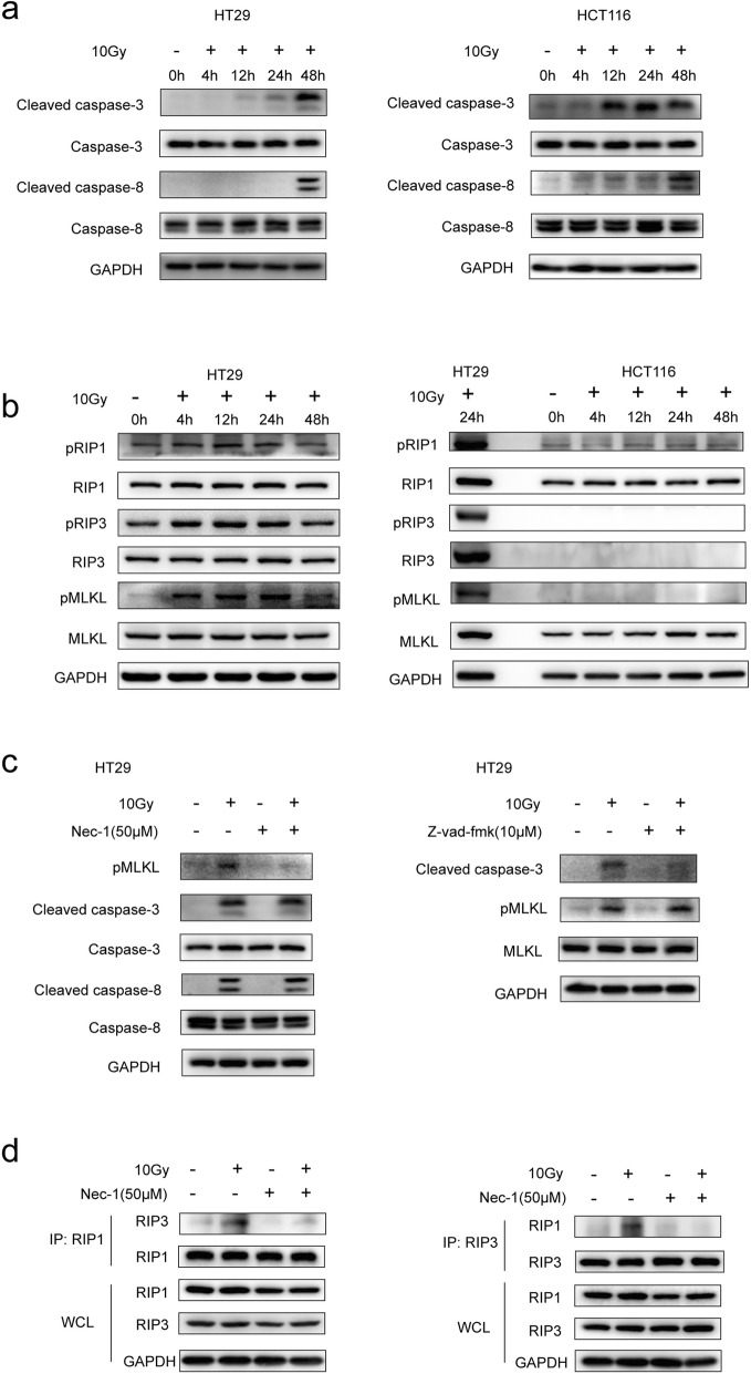
Fig. 1.
Western blot analysis of apoptosis and necroptosis associated proteins in colorectal cancer cell lines. a The full length and cleaved caspase-3 and 8 level at different time points post 10Gy irradiation in HT29 and HCT116 cells were evaluated by western blot. b The RIP1/pRIP1 (S166), RIP3/pRIP3(S227), MLKL/pMLKL(T357/S358) level at different time points post 10Gy irradiation in HT29 and HCT116 cells were evaluated by western blot. c Left panel, pMLKL, full length and cleaved caspase-3 and 8 level in HT29 cells at 48 h post 10Gy irradiation and pre-treatment with or without 50 μm Nec-1. Right panel, the cleaved caspase-3 and MLKL/pMLKL in HT29 cells at 24 h post 10Gy irradiation and pre-treatment with or without 10 μm Z-vad-fmk. d Co- immunoprecipitation of RIP1/RIP3 showed the formation of RIP1/RIP3 complex in irradiated HT29 cells and pre-treatment with or without 50 μm Nec-1. The endogenous RIP1 and RIP3 expression were determined using whole-cell lysates (WCL)

