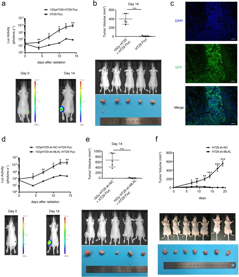
Fig. 5.
Knockdown of MLKL inhibited the growth stimulation effect and tumorigenicity in vivo. a–c Effect of dying HT29 on growth of HT29 Fluc in vivo. a Upper panel, proliferation of HT29 Fluc in vivo was monitored by bioluminescence imaging, student’s t-test, n = 5, lower panel, representative bioluminescent images of mice on Day 0 and Day 14. b Tumor volume of mice (upper panel) and photograph of xenograft (lower panel) on Day 14, student’s t-test, n = 5. c Immunofluorescence analysis showed GFP expression in HT29 tumor. Scale bar: 50 μm. d, e Effect of MLKL knockdown in dying HT29 cells on growth of HT29 Fluc in vivo. d Upper panel, proliferation of HT29 Fluc in vivo was monitored by bioluminescence imaging, student’s t-test, n = 5. Lower panel, representative bioluminescent images of mice on Day 0 and Day 14. e Tumor volume of mice (upper panel) and photograph of xenograft (lower panel) on Day 14, student’s t-test, n = 5. f Upper panel, xenograft tumor growth in nude mice from HT29 vector-transfected cells and HT29 sh-MLKL cells, student’s t-test, n = 7. Lower panel, Photograph of nude mice bearing HT29 vector-transfected and HT29 sh-MLKL xenograft. * < p 0.05, ** p < 0.005, *** p < 0.001

