Fig. 3.
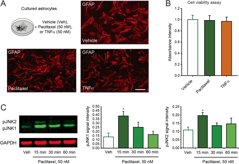
Paclitaxel induces the activation of cultured astrocytes. a Schematic illustration of the experimental conditions and immunofluorescence of GFAP in cultured astrocytes 24 h after exposure to vehicle control, paclitaxel, or TNF-α. Scale bar = 20 μm. b Quantification of astrocyte viability 24 h after incubation with vehicle control, paclitaxel, or TNF-α. c Western blot showing the phosphorylation/activation of JNK in cultured astrocytes after paclitaxel, as well as density of pJNK1 and pJNK2 bands, which are normalized to and expressed as ratio of the GAPDH loading control (*P < 0.05 compared to vehicle, ANOVA, n = 4 per group)

