Fig. 4.
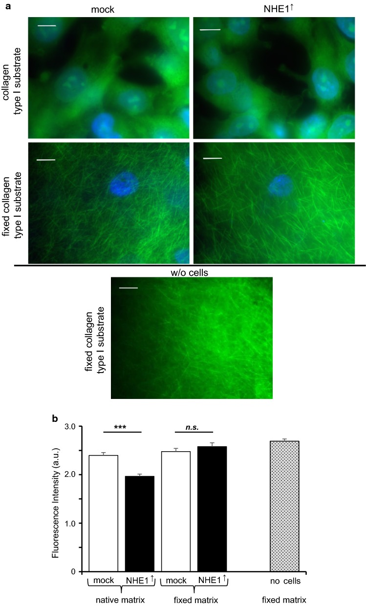
Digestion of collagen type I is fostered by NHE1 expression. a Fluorescence images of MV3 cells on native (upper images) and fixed collagen type I substrate (second row). The lowest image shows a fixed collagen type I substrate without (w/o) cells. Cells were kept on the respective substrate for 48 h and then fixed with glutaraldehyde. While the fixed substrate is characterized by a network of collagen I fibers the native substrate has been remodeled and mostly digested. Blue: DAPI staining of nuclei; green: glutaraldehyde-induced autofluorescence. Scale bar: 10 µm. b Fluorescence intensity measurements of native and fixed matrices. Native matrices populated with NHE1 overexpressing MV3 cells (n = 49 areas from N = 4 independent experiments) show a significantly lower intensity than those with control cells (n = 25, N = 2). Regardless of whether being populated with NHE1 overexpressing (n = 40, N = 9) or control cells (n = 15, N = 3), the intensities of the fixed substrates did not differ from that of unsettled matrices (n = 18, N = 3)

