Fig. 4.
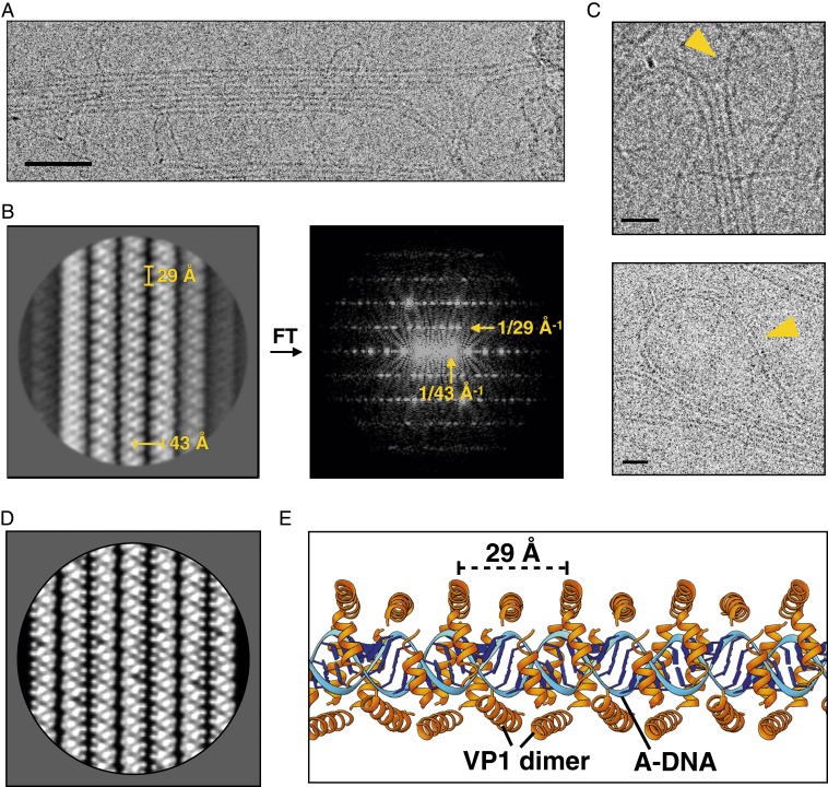
Cryo-EM of the disrupted SPV1 nucleoprotein genome. (A) Electron micrograph of extracted SPV1 genome shows filaments bundled into 2D rafts. (Scale bar, 500 Å.) (B) Two-dimensional class average of rafts (Left) and its power spectrum (Right) shows the 29-Å VP1-dimer axial repeat and the 43-Å interfilament spacing. (C) Electron micrographs show the adjacent filaments within the same raft connected by loops. (Scale bars, 200 Å.) (D) Two-dimensional raft projection generated from a VP1-DNA filament model, filtered to a 10-Å resolution. (E) A model for the nucleoprotein filament showing A-form DNA (blue) and VP1 dimers (orange) wrapped around the DNA.

