Fig. 5.
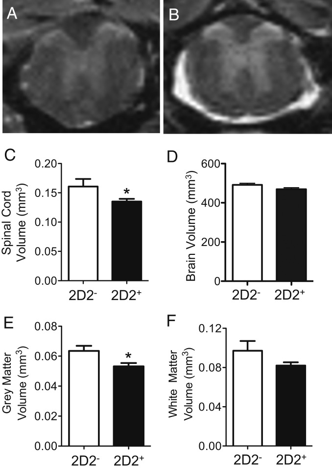
Spinal cord atrophy is prominent in PPARαmut/WT 2D2+ mice with age as detected by MR. PPARαmut/WT 2D2+ females and sex-matched PPARαmut/WT 2D2− littermates, aged 9 mo (n = 5 mice per group), were killed, and the spinal cord and brain specimens were fixed and imaged by MR. (A and B) Examples of MR images of the spinal cord in aged 2D2− or 2D2+ mice. (C–F) Volumes of the whole spinal cord (C), the whole brain (D), spinal cord gray matter (E), and spinal cord white matter (F) in 2D2+ and 2D2− mice. Means + SEM. *Significantly different from 2D2− mice by Mann–Whitney U test (2-tailed) (P ≤ 0.05).

