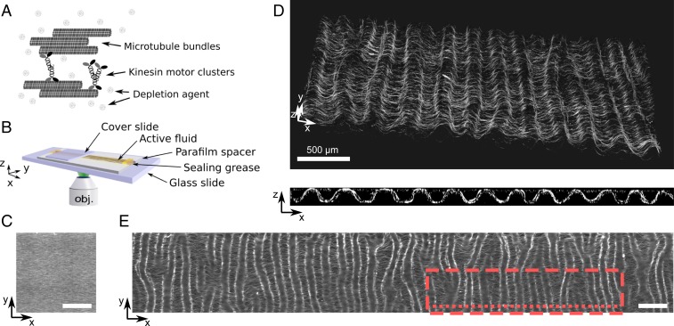
Fig. 1.
At low motor concentration a 3D active nematic fluid creates a thin corrugated sheet of well-defined wavelength. (A) Scheme of the components of the active fluid formed by nongrowing microtubules bundled together by a depletion agent and clusters of kinesin motors. (B) Scheme of the channel where the fluid (in yellow) is observed. (C) Epifluorescence image of the fluid at initial time. (D) Confocal images in 3D (Top) and cross-section in the plane (Bottom) of the fluid after 300 min. (E) Epifluorescence image of the same sample after 24 h and over a - area. The red dashed rectangle and the red dotted line respectively indicate the region where Top and Bottom images in D were recorded. (Scale bars, 500 m; motor concentration, 0.5 nM.)

