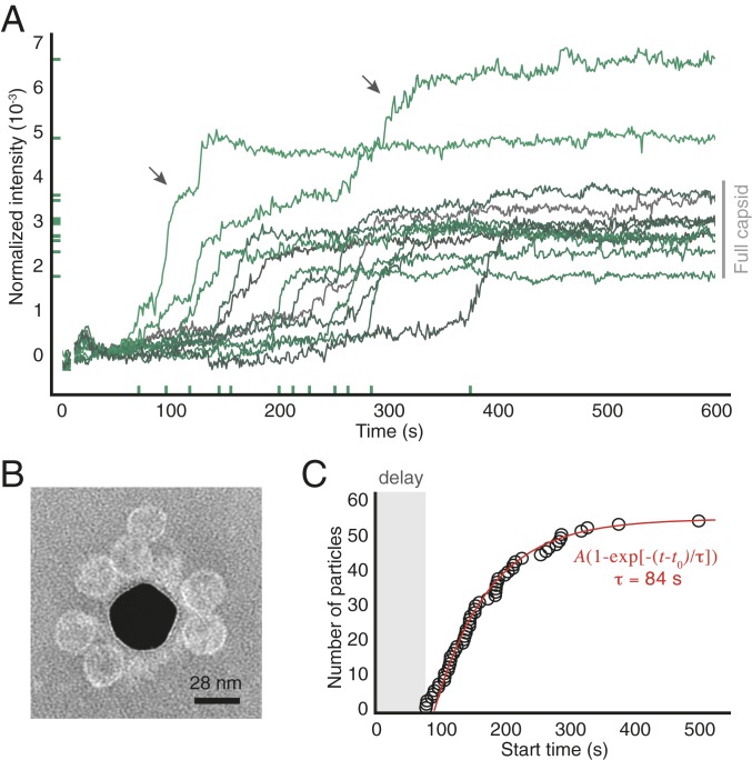
Fig. 2.
Assembly of 2-M coat-protein dimers around surface-tethered RNA strands. (A) Intensity traces for 12 randomly chosen particles from one experiment. x-axis ticks show the start times and y-axis ticks the final intensities. Gray bar indicates the intensity range corresponding to wild-type capsids. Arrows show 2 traces corresponding to overgrown particles. (B) Negatively stained TEM image of particles assembled around RNA strands tethered to a gold nanoparticle (dark region at center). We use a nanoparticle as the substrate because TEM cannot image through a coverslip. (C) The cumulative distribution of start times of all of the traces in the experiment is well fitted by an exponential with delay time of 92 s and a characteristic time of 84 s (see SI Appendix for fit results from repeated experiments). Uncertainties in the start times are smaller than the diameter of the circles.

